


X-ray Diffraction (XRD) Facility
A research facility for investigation of single crystal and powder/thin film samples by X-Ray diffraction techniques.
RRID:SCR_022886
Training, experimental design and/or sample service.
Visit Us!
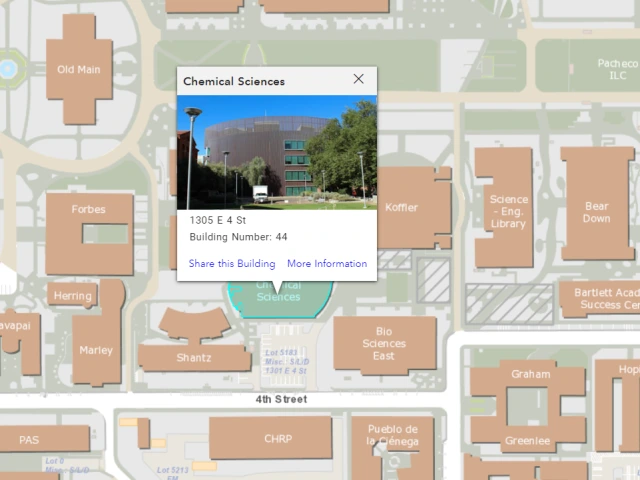
Location:
Chemical Sciences Bldg. (CSB), Room 117
Hours:
Monday thru Friday
8:00 AM to 5:00 PM
24/7 Access for Trained Users
Services
The XRD Facility serves the Faculty of the UA Department of Chemistry and outside customers, both academic and non-academic. We can use X-ray crystallography to determine the structure of small molecules by studying the XRD from single crystal samples. We can also study the powder and thin film samples to determine the crystallinity, composition, and various other properties of such samples (e.g., the thickness of thin films, preferred orientation of crystallites, etc.). The instrumentation available includes the single crystal X-ray diffractometers (Bruker D8 Venture Duo and Kappa Apex II Duo) and the powder diffractometer (Philips / Panalytical X’Pert Pro MPD). Contact the facility manager for details.
Education/Outreach
The facility performs, twice a year, lab demonstrations for students of Chem 412 class. The students willing to learn the use of the single crystal and/or powder XRD instruments are welcome. Such students should contact the manager to arrange for training.
Facility Publications
Use the Keck RRID!
University of Arizona - CBC X-Ray Diffraction (XRD) Facility, RRID:SCR_022886
All experiments on the Bruker D8 Venture Duo must include the following in the acknowledgements section: "The Bruker D8 Venture Duo instrument purchase was supported by the National Science Foundation under Grant Number CHE 2117516."
PUBLICATIONS 2022
Ahn, J; Wu, J; Ahn, J; Lee, J. Arsenic trioxide leaching and scorodite crystallization in methanesulfonic acid. Miner. Process. Extr. Metall. Rev. 43 (2022) 969-977.
Ahn, J; Wu, J; Lee, J. A Comparative Kinetic Study of Chalcopyrite Leaching Using Alternative Oxidants in Methanesulfonic Acid System. Miner. Process. Extr. Metall. Rev. 43 (2022), 390-401.
Encinas-Basurto, D; Konhilas, JP; Polt, R; Hay, M; Mansour, HM. Glycosylated Ang-(1-7) MasR Agonist Peptide PolyLactic-co-Glycolic Acid (PLGA) Nanoparticles and Microparticles in Cognitive Impairment: Design, Particle Preparation, Physicochemical Characterization, and In Vitro Release. Pharmaceutics 14 (2022) 587.
Keersmaecker, M De; Armstrong, NR; Ratcliff, EL. "How Low Can You Go? Defect Quantification at the 1015 cm–3 Level in Mixed-Cation Perovskites Using Differential Pulse Voltammetry." ACS Energy Lett. 2022, 7 4017-4027.
Rios-Valenciana, EE; Menezes, O; Niu, XZ; Romero, J; Root, RA; Chorover, J; Sierra-Alvarez, R; Field, JA. Reductive transformation of the insensitive munitions compound nitroguanidine by different iron-based reactive minerals. Environ. Pollut. 309 (2022) 119788.
Rushlow, J; Astashkin, AV; Albert, DR; Rajaseelan, E. “[(1,2,5,6-η)-Cycloocta-1,5-diene](4-isopropyl-1-methyl-1,2,4-triazol-5-ylidene)(tricyclohexylphosphane-κP)iridium(I) tetrafluoridoborate dichloromethane monosolvate,” IUCrData 7 (2022) x220685.
Vallorz, EL; Encinas-Basurto, D; Schnellmann, RG; Mansour, HM. Design, Development, Physicochemical Characterization, and In Vitro Drug Release of Formoterol PEGylated PLGA Polymeric Nanoparticles. Pharmaceutics 14 (2022) 638.
Wang, YC; Kegel, LL; Knoff, DS; Deodhar, BS; Astashkin, AV; Kim, M; Pemberton, JE. "Layered supramolecular hydrogels from thioglycosides." J. Mater. Chem B 2022, 10 3861-3875.
Wu, W.; Sung, Y.-S.; Tomat, E. “Thiol-Reactive Arylsulfonate Masks for Phenolate Donors in Antiproliferative Iron Prochelators” Inorg. Chem. 2022, 61, 49, 12457-12466.
PUBLICATIONS 2021
Acosta MF, Muralidharan P, Grijalva CL, Abrahamson MD, Hayes D Jr, Fineman JR, Black SM, Mansour HM. Advanced therapeutic inhalation aerosols of a Nrf2 activator and RhoA/Rho kinase (ROCK) inhibitor for targeted pulmonary drug delivery in pulmonary hypertension: design, characterization, aerosolization, in vitro 2D/3D human lung cell cultures, and in vivo efficacy. Ther. Adv. Respir. Dis. 15 (2021) 1–25.
Alabsi W, Acosta MF, Al-Obeidi FA, Hay M, Polt R, Mansour HM. Synthesis, Physicochemical Characterization, In Vitro 2D/3D Human Cell Culture, and In Vitro Aerosol Dispersion Performance of Advanced Spray Dried and Co-Spray Dried Angiotensin (1-7) Peptide and PNA5 with Trehalose as Microparticles/Nanoparticles for Targeted Respiratory Delivery as Dry Powder Inhalers. Pharmaceutics 13 (2021) 1278.
Alabsi W, Al-Obeidi FA, Polt R, Mansour HM. Organic Solution Advanced Spray-Dried Microparticulate/Nanoparticulate Dry Powders of Lactomorphin for Respiratory Delivery: Physicochemical Characterization, In Vitro Aerosol Dispersion, and Cellular Studies. Pharmaceutics 13 (2021) 26.
Castaldi, KT; Astashkin, AV; Albert, DR; Rajaseelan, E. “(4-benzyl-1-methyl-1,2,4-triazol-5-ylidene)[(1,2,5,6-η)-Cycloocta-1,5-diene](triphenylphosphane)iridium(I)tetrafluoridoborate,” IUCrData 6 (2021) x211142.
Curtis, C. J.; Astashkin, A. V.; Conradie, J.; Ghosh, A.; Tomat, E. “Ligand-centered triplet diradical supported by a binuclear palladium(II) dipyrrindione” Inorg. Chem. 2021, 60, 12457-12466.
Delost, M. D.; Njardarson, J. T. “Mild Darzens Annulations for Assembly of Trifluorothiomethylated (SCF3) Aziridine and Cyclopropane Structures” Org. Lett. 2021, 23, 6121-6125.
Das, P.; Delost, M. D.; Qureshi, M. H.; Bao, J.; Fell, J. S.; Houk, K. N.; Njardarson, J. T. “Dramatic Effect of g-Heteroatom Dienolate Substituents on Counterion Assisted Asymmetric Anionic Amino-Cope Reaction Cascades” J. Am. Chem. Soc. 2021, 143, 5793-5804.
Dubrovin, VD; Zhu, X; Mollaee, M; Zong, J; Peyghambarian, N. Highly Dy2O3 and Er2O3 doped boron-aluminosilicate glasses for magneto-optical devices operating at 2 µm. J. Non Cryst. Solids 569 (2021) 120986.
Jariwala, S; Burke, S; Dunfield, S; Shallcross, RC; Taddei, M; Wang, J; Eperon, GE; Armstrong, NR; Berry, JJ; Ginger, DS. "Reducing surface recombination velocity of methylammonium-free mixed-cation mixed-halide perovskites via surface passivation." Chem. Mater. 2021, 33, 5035-5044.
Keersmaecker, M De; Armstrong, NR; Ratcliff, EL. "Defect quantification in metal halide perovskites: the solid-state electrochemical alternative." Energy Environ. Sci. 2021, 14, 4840-4846.
Lauta, N. R.; Williams, R. E.; Smith, D. T.; Njardarson, J. T. “New Oxidative Route to Indoles via Amino-Hydroxylation of ortho-Allenyl Anilines” J. Org. Chem. 2021, 86, 10713-10723.
Menezes, O; Yu, Y; Root, RA; Gavazza, S; Chorover, J; Sierra-Alvarez, R; Field, JA. Iron (II) monosulfide (FeS) minerals reductively transform the insensitive munitions compounds 2, 4-dinitroanisole (DNAN) and 3-nitro-1, 2, 4-triazol-5-one (NTO). Chemosphere 285 (2021) 131409
Muralidharan P, Acosta MF, Gomez AI, Grijalva C, Tang H, Yuan JX, Mansour HM. Design and Comprehensive Characterization of Tetramethylpyrazine (TMP) for Targeted Lung Delivery as Inhalation Aerosols in Pulmonary Hypertension (PH): In Vitro Human Lung Cell Culture and In Vivo Efficacy. Antioxidants (Basel) 10 (2021) 427.
Muralidharan P, Hayes D Jr, Fineman JR, Black SM, Mansour HM. Advanced Microparticulate/Nanoparticulate Respirable Dry Powders of a Selective RhoA/Rho Kinase (Rock) Inhibitor for Targeted Pulmonary Inhalation Aerosol Delivery. Pharmaceutics. 13 (2021) 2188.
Newman, EB; Astashkin, AV; Albert, DR; Rajaseelan, E. “(4-benzyl-1-methyl-1,2,4-triazol-5-ylidene)-[(1,2,5,6-η)-Cycloocta-1,5-diene](triphenylphosphane)iridium(I) tetrafluoridoborate,” IUCrData 6 (2021) x210836.
Rao, PR; Muralidharan, K; Momayez, M; Runge, K. A multiscale microstructural characterization of SiO2-Al2O3 foams. Mater. Charact. 181 (2021) 111433.
Rood, JA; Subedi, CB; Risell, JP; Astashkin, AV; Rajaseelan, E. “[(1,2,5,6-η)-Cycloocta-1,5-diene](1-ethyl-3-isopropyl-1,3-imidazol-2-ylidene)(triphenylphosphane)rhodium(I) tetrafluoridoborate,” IUCrData 6 (2021) x210597.
Rushlow, J; Astashkin, AV; Albert, DR; Rajaseelan, E. “(Chlorido/bromido)[(1,2,5,6-η)-cycloocta-1,5-diene](4-isopropyl-1-methyl-1,2,4-triazol-5-ylidene)rhodium(I),” IUCrData 6 (2021) x210811.
Scott, K. A.; Groch, J. R.; Chogii, I.; Delost, M. D.; Das, P.; Njardarson, J. T. “Dienolate Annulation Approach for Assembly of Densely Substituted Aromatic Architectures” J. Org. Chem. 2021, 86, 10555-10555.
Shaikh, A.C.; Veleta, J.M.; Moutet, J; Gianetti, T.L. T"rioxatriangulenium (TOTA+) as a robust carbon-based Lewis acid in frustrated Lewis pair chemistry." Chem. Sci. 2021, 12, 4841-4849.
Shallcross, RC; Armstrong, NR. "Near-Surface Composition, Structure, and Energetics of TiO2 Thin Films: Characterization of Stress-Induced Defect States in Oxides Prepared via Chemical Vapor Deposition versus Solution Deposition from Sol–Gel Precursors." J. Phys. Chem. C 2021, 125, 24011-24024.
Shepard, AJ; Townsend, JA; Foley, C; Hulme, C; Marty, MT; Jewett, JC. Suzuki Coupling of Protected Aryl Diazonium Ions: Expanding the Knowledge of Triazabutadiene Compatible Reactions. Org. Lett. 23 (2021) 1851-1855.
Sung, Y.-S.; Wu, W.; Ewbank, M. A.; Utterback, R. D.; Marty, M. T.; Tomat, E. “Albumin conjugates of thiosemicarbazone and imidazole-2-thione prochelators: Iron coordination and antiproliferative activity” ChemMedChem 2021, 16, 2764-2768.
Tomat, E., Curtis, C. J. “Biopyrrin pigments: From heme metabolites to redox-active ligands and luminescent radicals” Acc. Chem. Res. 2021, 54, 4584-4594.
Zhang, Y; Ji, P; Gao, F; Dong, Y; Huang, H; Wang, C; Zhou, Z; Wang, W. "Organophotocatalytic dearomatization of indoles, pyrroles and benzo (thio) furans via a Giese-type transformation." Commun. Chem. 2021, 4, 1-13.
Zhang, Y; Ji, P; Gao, F; Huang, H; Zeng, F; Wang, W. "Photoredox Asymmetric Nucleophilic Dearomatization of Indoles with Neutral Radicals." ACS Catal. 2021, 11, 998-1007.
PUBLICATIONS 2020
Acosta MF, Muralidhran P, Abrahamson MD, Grijalva CL, Carver M, Tang H, Klinger C, Fineman JR, Black SM, Mansour HM. Comparison of l-Carnitine and l-Carnitine HCL salt for targeted lung treatment of pulmonary hypertension (PH) as inhalation aerosols: Design, comprehensive characterization, in vitro 2D/3D cell cultures, and in vivo MCT-Rat model of PH. Pulm. Pharmacol. Ther. 65 (2020) 101998.
Acosta MF, Abrahamson MD, Encinas-Basurto D, Fineman JR, Black SM, Mansour HM. Inhalable Nanoparticles/Microparticles of an AMPK and Nrf2 Activator for Targeted Pulmonary Drug Delivery as Dry Powder Inhalers. AAPS J. 23 (2020) 2.
Astashkin, A. V.; Utterback, R. D.; Sung, Y.-S.; Tomat, E. “Iron complexes of an antiproliferative aroyl hydrazone: Characterization of three protonation states by EPR methods” Inorg. Chem. 2020, 59, 11377-11384.
Curtis, C. J.; Tomat, E. “Heteroleptic palladium(II) complexes of dipyrrin-1,9-dione supported by intramolecular hydrogen bonding” J. Porphyrins Phthalocyanines 2020, 24, 112-120.
Delost, M. D.; Njardarson, J. T. “Strategic Vinyl Sulfone Nucleophile b-Substitution Significantly Impacts Selectivity in Vinylogous Darzens and aza-Darzens Reactions” Org. Lett. 2020, 22, 6917.
Gomez AI, Acosta MF, Muralidharan P, Yuan JX, Black SM, Hayes D Jr, Mansour HM. Advanced spray dried proliposomes of amphotericin B lung surfactant-mimic phospholipid microparticles/nanoparticles as dry powder inhalers for targeted pulmonary drug delivery. Pulm Pharmacol Ther. 64 (2020) 101975.
Karayilan, M.; McCleary-Petersen, K. C.; Hamilton, M. O’B.; Fu, L.; Matyjaszewski, K.; Glass, R. S.; Lichtenberger, D. L.; Pyun, J. Polymerization from a [2Fe-2S] Metalloinitiator: Molecular Weight Effects on Electrocatalytic Hydrogen Production. Macromol. Rapid Commun. 2020, 41(1), 1900424.
Ma, D; Ngo, V; Raghavan, S; Sandoval, S. Degradation of Ir-Ta oxide coated Ti anodes in sulfuric acid solutions containing fluoride. Corros. Sci. 164 (2020) 108358.
Muralidharan P, Mallory EK, Malapit M, Phan H, Ledford JG, Hayes D Jr, Mansour HM. Advanced design and development of nanoparticle/microparticle dual-drug combination lactose carrier-free dry powder inhalation aerosols. RSC Adv. 10 (2020) 41846-41856.
Muralidharan P, Jones B, Allaway G, Biswal SS, Mansour HM. Design and development of innovative microparticulate/nanoparticulate inhalable dry powders of a novel synthetic trifluorinated chalcone derivative and Nrf2 agonist. Sci Rep. 10 (2020) 19771.
Rao, PR; Muralidharan, K; Momayez, M; Runge, KA; Loy, DA. Direct foaming driven synthesis and thermophysical characterization of silica-alumina foams: Applications for thermal insulation. Ceram. Int. 46 (2020), 10431-10441.
Schofield, K; Foley, C; Hulme, C. 5-Endo Trig Oxidative Radical Cyclizations of Ugi-3CR Products toward 1,4-Imidazolidinones. Org. Lett. 23 (2020) 107-112.
PUBLICATIONS 2019
Ahn, J; Wu, J; Ahn, J; Lee, J. Comparative investigations on sulfidic gold ore processing: A novel biooxidation process option. Miner. Eng. 140 (2019) 105864.
Chogii, I.; Das, P.; Njardarson, J. T. “Efforts toward a Unified Kainoid Family Synthesis Approach: Unexpected Sulfinamide Directed Conjugate Addition Results”, Asian. J. Org. Chem. 2019, 8, 1041-1044.
Gautam, R.; Petritis, S. J.; Tomat, E. “Redox-switchable cyan fluorescence of a BODIPY analog inspired by propentdyopent pigments” Eur. J. Inorg. Chem. 2019, 68-72.
Glass, R. S.; Pyun, J.; Lichtenberger, D. L.; Brezinski, W. P.; Karayilan, M.; Clary, K. E.; Pavlopoulos, N. G.; Evans, D. H. Water-soluble and air-stable [2Fe-2S]-metallopolymers: A new class of electrocatalysts for H2 production via water splitting. Phosphorus Sulfur Silicon Relat. Elem. 2019, 194(7), 701-706.
Shallcross, RC; Olthof, S; Meerholz, K; Armstrong, NR. "Impact of titanium dioxide surface defects on the interfacial composition and energetics of evaporated perovskite active layers." ACS Appl. Mater. Interfaces 2019, 11, 32500-32508.
Tomat, E. “Propentdyopents: Brief history of a family of dipyrrolic pigments" J. Porphyrins Phthalocyanines 2019, 23, 1265-1272.
PUBLICATIONS 2018
Brezinski, W. P.; Karayilan, M.; Clary, K. E.; McCleary-Petersen, K. C.; Fu, L.; Matyjaszewski, K.; Evans, D. H.; Lichtenberger, D. L.; Glass, R. S.; Pyun, J. Macromolecular Engineering of the Outer Coordination Sphere of [2Fe-2S] Metallopolymers to Enhance Catalytic Activity for H2 Production. ACS Macro Lett. 2018, 7(11), 1383-1387.
Brezinski, W. P.; Karayilan, M.; Clary, K. E.; Pavlopoulos, N. G.; Li, S.; Fu, L.; Matyjaszewski, K.; Evans, D. H.; Glass, R. S.; Lichtenberger, D. L.; Pyun, J. [FeFe]-hydrogenase mimetic metallopolymers with enhanced catalytic activity for hydrogen production in water. Angew. Chem. Int. Ed. 2018, 57, 11898-11902.
Chogii, I.; Das, P.; Delost M. D.; Crawford, M. N.; Njardarson, J. T. “Asymmetric Vinylogous aza-Darzens Approach to Vinyl Aziridines”, Org. Lett. 2018, 20, 4942-4945.
Smith, B. R.; Njardarson, J. T. “[2.2.2] to [3.2.1]-Bicycle Rearrangement Approach to the Gibberellin Family of Natural Products”, Org. Lett. 2018, 20,2993-2996.
Publications 2017
Idrees, KB; Rutledge, WJ; Roberts, SA; Rajaseelan, E. “[(1,2,5,6-η)-1,5-Cyclooctadiene] (1-ethyl-3-isopropyl-1,3-imidazol-2-ylidene(triphenylphosphine)iridium(I)Tetrafluoroborate,” IUCrData 2 (2017) x171411.
Idrees, KB; Astashkin, AV; Rajaseelan, E. "[µ-1,4-Bis(diphenylphosphine)butane] bis{(4-benzyl-2-neopentylo-1,2,4-triazol-3-ylidene)[1,2,5,6-η)-1,5-Cyclooctadiene]iridium(I)} bis(tetrafluoroborate) dicholoromethane disolvate,” IUCrData 2 (2017) x171081.
Martinez-Ariza, G; Mehari, BT; Pinho, LAG; Foley, C; Day, K; Jewett, JC; Hulme, C. Synthesis of fluorescent heterocycles via a Knoevenagel/[4+ 1]-cycloaddition cascade using acetyl cyanide. Org. Biomol. Chem. 15 (2017) 6076-6079.
Weisbart, C; Raghavan, S; Muralidharan, K; Potter Jr, BG. Electrocoagulation driven fabrication of graphene oxide films. Carbon 116 (2017) 318-324.
PUBLICATIONS 2011
Little EJ, Dunten PW, Bitinaite J, Horton NC. New clues in the allosteric activation of DNA cleavage by SgrAI: structures of SgrAI bound to cleaved primary-site DNA and uncleaved secondary-site DNA. Acta Crystallogr D: Biol. Crystallogr. 2011; 67, 67-74.
PUBLICATIONS 2010
Park CK, Joshi HK, Agrawal A, Ghare MI, Little EJ, Dunten PW, Bitinaite J, Horton NC. Domain swapping in allosteric modulation of DNA specificity PLOS Biol. 2010; 8, e1000554.
Horton NC, Park CK. Crystallization of zinc finger proteins bound to DNA. Methods Mol Biol. 2010; 649:457-77.
Weichsel A, Kem M, Montfort WR. Crystal structure of human thioredoxin revealing an unraveled helix and exposed S-nitrosation site.Protein Sci. 2010, 19 1801-1806.
PUBLICATIONS 2009
Robert E. Berry, Maxim N. Shokhirev, Arthur Y. W. Ho, Fei Yang, Tatiana K. Shokhireva, Hongjun Zhang, Andrzej Weichsel, William R. Montfort and F. Ann Walker. Effect of Mutation of Carboxyl Side-Chain Amino Acids Near the Heme on the Midpoint Potentials and Ligand Binding Constants of Nitrophorin 2 and Its NO, Histamine, and Imidazole Complexes J. Am. Chem. Soc., (2009), 131, 2313–2327.
Dunten PW, Little EJ, Horton NC.The restriction enzyme SgrAI: structure solution via combination of poor MIRAS and MR phases. Acta Crystallogr D Biol Crystallogr. (2009) , 65,393-8.
Campbell ZT, Weichsel A, Montfort WR, Baldwin TO. Crystal Structure of the Bacterial Luciferase/Flavin Complex Provides Insight into the Function of the β Subunit. Biochemistry, (2009), 48, 6085–6094.
Loftin IR, Blackburn NJ, McEvoy MM. Tryptophan Cu(I)-pi interaction fine-tunes the metal binding properties of the bacterial metallochaperone CusF. J Biol Inorg Chem. 2009, 14, 905-12.
Dexheimer TS, Carey SS, Zuohe S, Gokhale VM, Hu X, Murata LB, Maes EM, Weichsel A, Sun D, Meuillet EJ, Montfort, WR, Hurley LH. NM23-H2 may play an indirect role in transcriptional activation of c-myc gene expression but does not cleave the nuclease hypersensitive element III1. Mol Cancer Ther (2009), 8, 1363-1377.
PUBLICATIONS 2008
Matthew S. Dubrava, Wendy M. Ingram, Sue A. Roberts, Andrzej Weichsel, William R. Montfort, and Matthew H. J. Cordes . N15 Cro and λ Cro: orthologous DNA-binding domains with completely different but equally effective homodimer interfaces, Protein Science (2008), 17 , 803-812.
Shane D. Morrison, Sue A. Roberts, Abreeza M. Zegeer, William R. Montfort, and Vahe Bandarian. A new use for a familiar fold: The x-ray crystal structure of GTP-bound GTP cyclohydrolase III from Methanocaldococcus jannaschii reveals a two metal ion catalytic mechanism. Biochemistry (2008), 47, 230-242.
Hall, Branwen M., Roberts, Sue A., Heroux, Annie M., Cordes, Matthew H. Two structures of a λ-Cro variant highlight dimer flexibility but disfavor major dimer distortions upon specific binding of cognate DNA. J. Mol Biol . (2008), 375, 802-811.
Christian G. Roessler, Branwen M. Hall, William J. Anderson, Wendy M. Ingram, Sue A. Roberts, William R. Montfort, and Matthew H. J. Cordes. Transitive homology-guided structural studies lead to discovery of Cro proteins with 40% sequence identity but different folds. PNAS , (2008), 105, 2343-2348.
Pete W. Dunten, Elizabeth J. Little, Mark T. Gregory, Veena M. Manohar, Michael Dalton, David Hough, Jurate Bitinaite, and Nancy C. Horton. The structure of SgrAI bound to DNA; recognition of an 8 base pair target. Nucl. Acids Res. (2008), 36, 5405-5416.
Babic AC, Little EJ, Manohar VM, Bitinaite J, Horton NC. DNA distortion and specificity in a sequence-specific endonuclease. J Mol Biol. (2008), 383, 186-204.
James E. Spoonamore, Sue A. Roberts, Annie Heroux and Vahe Bandarian. X-ray crystal structure of a 6-pyruvoyltetrahydropterin synthase homolog from Streptomyces coelicolor . Acta Crystallogr. F. (2008), F64, 875-879.
Arvizu-Flores AA, Sugich-Miranda R, Arreola R, Garcia-Orozco KD, Velazquez-Contreras EF, Montfort WR, Maley F, Sotelo-Mundo RR. Role of an invariant lysine residue in folate binding on Escherichia coli thymidylate synthase: calorimetric and crystallographic analysis of the K48Q mutant. Int J Biochem Cell Biol. (2008), 40 2206-17.
Little EJ, Babic AC, Horton NC. Early interrogation and recognition of DNA sequence by indirect readout. Structure. (2008). 16, 1828-37.
PUBLICATIONS 2007
Shokhireva TKh, Weichsel A, Smith KM, Berry RE, Shokhirev NV, Balfour CA, Zhang H, Montfort WR, Walker FA. Assignment of the Ferriheme Resonances of the Low-Spin Complexes of Nitrophorins 1 and 4 by 1H and 13C NMR Spectroscopy: Comparison to Structural Data Obtained from X-ray Crystallography. Inorganic Chemistry, (2007), 46, 2041-2056.
Isabell R. Loftin, Sylvia Franke, Ninian J. Blackburn, and Megan M. McEvoy. Unusual Cu(I)/Ag(I) coordination of Escherichia coli CusF as revealed by atomic resolution crystallography and X-ray absorption spectroscopy Protein Sci, (2007), 16, 2287-2293.
Schreiter ER, Rodríguez MM, Weichsel A, Montfort WR, Bonaventura J. S-nitrosylation-induced conformational change in blackfin tuna myoglobin. J. Biol. Chem., (2007), 282, 19773-80.
Weichsel, A., Brailey, JL, Montfort, WR. Buried S-Nitrosocysteine Revealed in Crystal Structures of Human Thioredoxin. Biochemistry, (2007), 46, 1219-1227.
EARLIER PUBLICATIONS
Roberts SA, Hyatt, DC, Honts, JE, Changchien C, Maley, GF, Maley F, Montfort WR. Y94F Mutant of E. coli Thymidylate S ynthase. Acta Crystallogr F62 (2006), 840-843.
Segal DJ, Crotty JW, Bhakta MS, Barbas CF, Horton NC. Structure of Aart, a designed six-finger zinc finger peptide, bound to DNA. J Mol Biol. (2006), 363, 405-21.
Joshi HK, Etzkorn C, Chatwell L, Bitinaite J, Horton NC. Alteration of sequence specificity of the type II restriction endonuclease HincII through an indirect readout mechanism. J Biol Chem. (2006) 281,23852-69.
Weichsel A, Maes EM, Andersen JF, Valenzuela JG, Shokhireva, TK, Walker FA, and Montfort WR. Heme-assisted S-nitrosation of a proximal thiolate in a nitric oxide transport protein. Proc. Natl. Acad. Sci. USA, (2005), 102, 594-599.
Maes EM, Roberts SA, Weichsel A, Montfort WR. Heme Distortion in Nitrophorin 4: Ultra-High Resolution Structures of Ferrous Complexes. Biochemistry, (2005), 44,12690-12699.
Loftin IR, Franke S, Roberts SA, Weichsel A, Heroux A, Montfort WR, Rensing C, McEvoy M. A Novel Copper-binding Fold for the Periplasmic Metallochaperone Cusf. Biochemistry, (2005), 44, 10533-10540.
Berry RE, Ding XD, Shokhireva TK, Weichsel A, Montfort WR, Walker FA. Axial ligand complexes of the Rhodnius nitrophorins: reduction potentials, binding constants, EPR spectra, and structures of the 4-iodopyrazole and imidazole complexes of NP4. J Biol Inorg Chem. (2004), 9, 135-144.
Maes EM, Weichsel A, Andersen JF, Shepley D, Montfort WR. Role of Binding Site Loops in Controlling Nitric Oxide Release: Structure and Kinetics of Mutant Forms of Nitrophorin 4. Biochemistry (2004), 43, 6679-6690.
Kondrashov DA, Roberts SA, Weichsel A, Montfort WR. Protein functional cycle viewed at atomic resolution: conformational change and mobility in nitrophorin 4 as a function of pH and NO binding. Biochemistry (2004), 43, 13637-13647.
K. Nienhaus, E. M. Maes, A. Weichsel, W. R. Montfort and G. U. Nienhaus, Structural dynamics controls nitric oxide affinity in Nitrophorin 4 Journal of Biological Chemistry, (2004) 279, 39401-39407.
Roberts, Sue A.; Wildner, Guenter F.; Grass, Gregor; Weichsel, Andrzej; Ambrus, Attila; Rensing, Christopher; Montfort, William R. A Labile Regulatory Copper Ion Lies Near the T1 Copper Site in the Multicopper Oxidase CueO. J.Biol.Chem. (2003), 278, 31958-31963.
Roberts, Sue A.; Weichsel, Andrzej; Grass, Gregor; Thakali, Keshari; Hazzard, James T.; Tollin, Gordon; Rensing, Christopher; Montfort, William R. Crystal structure and electron transfer kinetics of CueO, a multicopper oxidase required for copper homeostasis in Escherichia coli. PNAS (2002), 99, 2766-2771.
Roberts, Sue A.; Weichsel, Andrzej; Qiu, Yan; Shelnutt, John A.; Walker, F. Ann; Montfort, William R. Ligand-Induced Heme Ruffling and Bent NO Geometry in Ultra-High-Resolution Structures of Nitrophorin 4. Biochemistry (2001), 40, 11327-11337.
Weichsel, Andrzej; Andersen, John F.; Roberts, Sue A.; Montfort, William R. Nitric oxide binding to nitrophorin 4 induces complete distal pocket burial. Nature Struct Biol. (2000), 7, 551-554.